Working as a multi-disciplinary collaboration, this H2020 project aims to combine the knowledge of leading researchers and pioneering industry specialists to hone the technology and expertise needed to develop ground-breaking ways of visualising gene transcription in living tissue. The genetic content of a cell determines its function. Genes are constantly switched on and off from the birth of the cell up until it dies. This highly dynamic process is regulated both by internal (genetic) and external (environmental) factors. In this H2020 funded project we develop a new technology to visualise the active genes in real time. In other words, we will be able to see when a gene is switched on and off in a living neuron.
CHEMISTRY
MICROSCOPY
SPECTROSCOPY
GENETICS
NEUROBIOLOGY
Dr John S. Fossey (University of Birmingham, UK) conducts the synthetic chemistry part of the project.
In the heart of the project is a novel high-tech microscope that is able to create images of activated genes.
The role of the Nanostructures and Applied Spectroscopy Group at Wigner RCP is to develop a tuneable stimulated Raman scattering (SRS) as the essential part of the novel microscope.
How DNA is transcribed and how we target genes of interest are covered here.
We are interested in how neurons work in health and diseases.
THE PROJECT
Observation and analysis of endogenous transcriptional processes in living and functioning organisms is not feasible today due to technical constraints. Therefore our understanding of the nuclear processes, and the options to diagnose or intervene at genetic level, is seriously constrained. Our proposal offers a cutting-edge technique to overcome this limitation. The aim is to visualize nuclear processes in intact brains in real-time, leading to: a new high-tech product development; prototyping of scientific equipment at the research institutes; and, in the long run, new tools to study and cure human diseases.
WELCOME TO THE NEURAM WEBSITE
The main objectives are to:
1) develop Stimulated Raman Spectroscopy (SRS)-based technologies to visualize endogenous transcription at single cell level;
2) develop tools for loading alkyne-tagged nucleosides and nucleotide analogues into neurons;
3) distinguish neuron types without labelling using SRS technology;
4) track transcription processes in intact brain with high temporal and spatial resolution;
5) develop and commercialise a 4D-SRS microscope for broad range of applications.
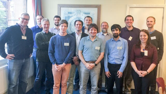
MEET THE TEAM
The ambition of NEURAM is reflected in the wide spread of interdisciplinary expertise of the consortium members. The consortium brings together leading academic and industrial partners, each of which has a distinct role or skill in the project.
Interdisciplinary project
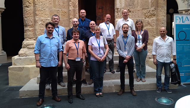
Summer school on Biophotonics
Complex projects cannot be carried out in a single lab as it require expertise in various fields. In this project we pulled together an international team from the field of chemistry, physics, neuroscience, nanobiology, genetics and interdisciplinary research management. This page briefly describes the expertise of each participant and the role in the project with links to the individual labs.
A successful summer school on Biophotonics (Photonics meets Biology) was organized with the Mesobrain H2020 project in Tarragona, Spain in September, 2017. All NEURAM consortium members participated with lectures.
In 2019 we co-organized and participated on the Photonics meets Biology summer school held in Crete, Greece. More info can be found here (http://esperia.iesl.forth.gr/~mfarsari/)
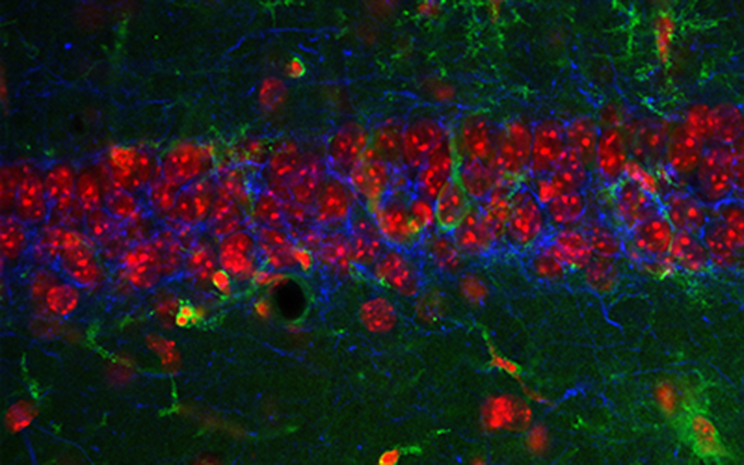
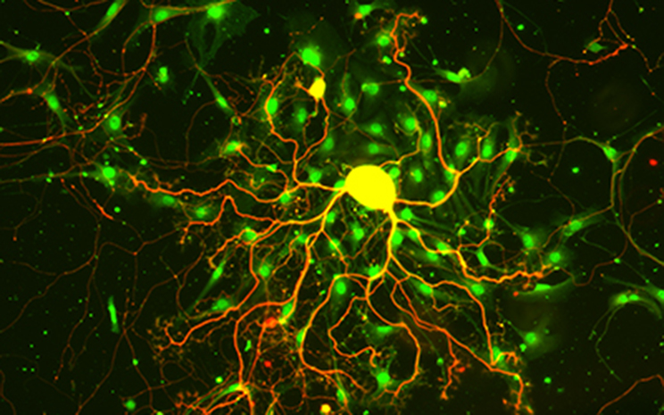
Successful magnefectation of mouse primary neuronal culture with FITC-labelled morpholinos. Mouse primary neuronal culture was culture for 5 days and then treated with magnetic nanoparticles (MNP) labelled with FITC-Morpholino, or non-labelled magnetic nanoparticles (Control) and magnefected for 30 minutes. Cells were fixed 1-day after treatment and observed on slides under a Zeiss confocal microscope. Blue colour represents DAPI stained nuclei, red colour shows the cell membrane and nanoparticles are labelled with green colour (FITC-morpholino).